
Rad21 antibody, Unconjugated, Rabbit, Polyclonal
Artikelnummer:
GTX106012
- Bilder (9)
| Artikelname: | Rad21 antibody, Unconjugated, Rabbit, Polyclonal |
| Artikelnummer: | GTX106012 |
| Hersteller Artikelnummer: | GTX106012 |
| Alternativnummer: | GTX106012-100,GTX106012-25 |
| Hersteller: | GeneTex |
| Wirt: | Rabbit |
| Kategorie: | Antikörper |
| Applikation: | ChIP, ICC, IHC-P, IP, WB |
| Spezies Reaktivität: | Human, Mouse, Rat |
| Immunogen: | Recombinant protein encompassing a sequence within the center region of human Rad21. The exact sequence is proprietary. |
| Konjugation: | Unconjugated |
| Alternative Synonym: | RAD21 cohesin complex component , CDLS4 , HR21 , HRAD21 , MCD1 , MGS , NXP1 , SCC1 , hHR21 |
| Anwendungsbeschreibung: | WB: 1:500-1:3000. ICC/IF: 1:100-1:1000. IHC-P: 1:100-1:1000. IP: 1:100-1:500. *Optimal dilutions/concentrations should be determined by the researcher.Not tested in other applications. |
|
|
GTX106012 IHC-P Image |
|
|
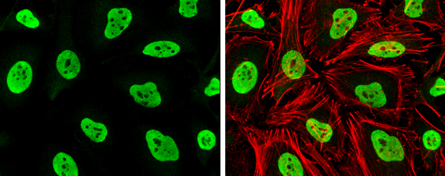
Rad21 antibody detects Rad21 protein at nucleus by immunofluorescent analysis.Sample: HeLa cells were fixed in 4% paraformaldehyde at RT for 15 min.Green: Rad21 stained by Rad21 antibody (GTX106012) diluted at 1:1000.Red: phalloidin, a cytoskeleton marker, diluted at 1:100. |
|
|
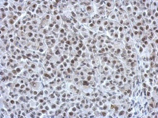
Immunohistochemical analysis of paraffin-embedded HBL435 xenograft, using RAD21(GTX106012) antibody at 1:750 dilution. Antigen Retrieval: Trilogy™ (EDTA based, pH 8.0) buffer, 15min |
|
|
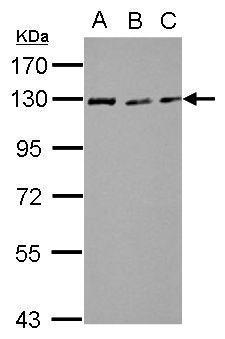
Sample (30 μg of whole cell lysate) A: NIH-3T3 B: JC C: BCL-1 7.5% SDS PAGE GTX106012 diluted at 1:3000 The HRP-conjugated anti-rabbit IgG antibody (GTX213110-01) was used to detect the primary antibody. |
|
|
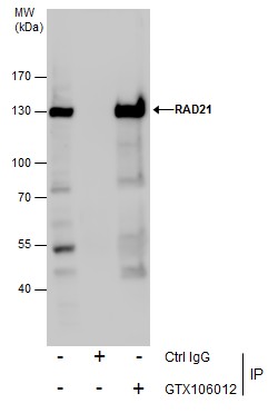
Immunoprecipitation of RAD21 protein from Jurkat whole cell extracts using 5 μg of RAD21 antibody (GTX106012). Western blot analysis was performed using RAD21 antibody (GTX106012). EasyBlot anti-Rabbit IgG (GTX221666-01) was used as a secondary reagent. |
|
|
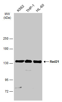
Various whole cell extracts (30 μg) were separated by 5% SDS-PAGE, and the membrane was blotted with Rad21 antibody (GTX106012) diluted at 1:1000. The HRP-conjugated anti-rabbit IgG antibody (GTX213110-01) was used to detect the primary antibody. |
|
|
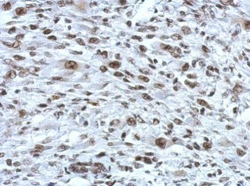
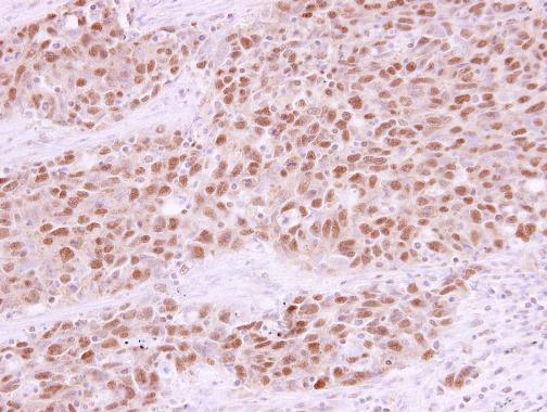
RAD21 antibody detects RAD21 protein at nucleus in human lung papillary adenocarcinoma by immunohistochemical analysis. Sample: Paraffin-embedded human lung papillary adenocarcinoma. RAD21 antibody (GTX106012) diluted at 1:250. |
|
|
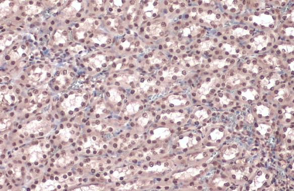
Rad21 antibody detects Rad21 protein at nucleus by immunohistochemical analysis.Sample: Paraffin-embedded rat kidney.Rad21 stained by Rad21 antibody (GTX106012) diluted at 1:500.Antigen Retrieval: Citrate buffer, pH 6.0, 15 min |
|
|
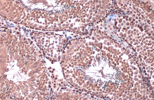
Rad21 antibody detects Rad21 protein at nucleus by immunohistochemical analysis.Sample: Paraffin-embedded mouse testis.Rad21 stained by Rad21 antibody (GTX106012) diluted at 1:500.Antigen Retrieval: Citrate buffer, pH 6.0, 15 min |
Produktgarantie und fachkundiger Support
