
TDP43 antibody, Unconjugated, Rabbit, Polyclonal
Artikelnummer:
GTX114210
- Bilder (10)
| Artikelname: | TDP43 antibody, Unconjugated, Rabbit, Polyclonal |
| Artikelnummer: | GTX114210 |
| Hersteller Artikelnummer: | GTX114210 |
| Alternativnummer: | GTX114210-100,GTX114210-25 |
| Hersteller: | GeneTex |
| Wirt: | Rabbit |
| Kategorie: | Antikörper |
| Applikation: | ChIP, ICC, IHC-P, IP, WB |
| Spezies Reaktivität: | Human, Mouse, Rat |
| Immunogen: | Recombinant protein encompassing a sequence within the center region of human TDP43. The exact sequence is proprietary. |
| Konjugation: | Unconjugated |
| Alternative Synonym: | TAR DNA binding protein , ALS10 , TDP-43 |
| Anwendungsbeschreibung: | WB: 1:500-1:3000. ICC/IF: 1:100-1:1000. IHC-P: 1:100-1:1000. IP: 1:100-1:1000. *Optimal dilutions/concentrations should be determined by the researcher.Not tested in other applications. |
|
|
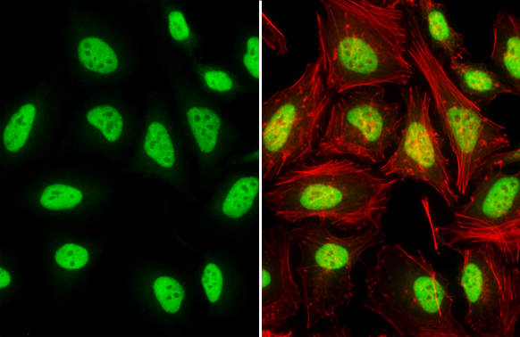
GTX114210 ICC/IF Image |
|
|
TDP43 antibody detects TDP43 protein by immunofluorescent analysis.Sample: DIV10 rat E18 primary cortical neuron cells were fixed in 4% paraformaldehyde at RT for 15 min.Green: TDP43 stained by TDP43 antibody (GTX114210) diluted at 1:500.Red: Tau, stained by Tau antibody [GT287] (GTX634809) diluted at 1:500.Blue: Fluoroshield with DAPI (GTX30920). |
|
|
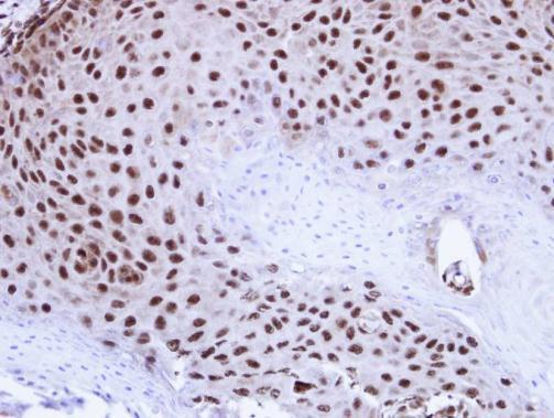
Immunohistochemical analysis of paraffin-embedded Cal27 Xenograft , using TDP-43(GTX114210) antibody at 1:100 dilution. Antigen Retrieval: Trilogy™ (EDTA based, pH 8.0) buffer, 15min |
|
|
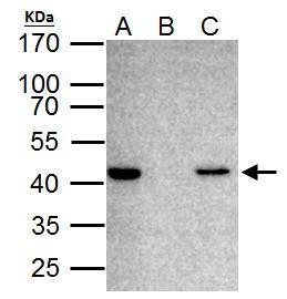
TARDBP antibody immunoprecipitates TARDBP protein in IP experiments. IP Sample: HeLa whole cell lysate/extract A. 40 μg HeLa whole cell lysate/extract B. Control with 2 μg of preimmune rabbit IgG C. Immunoprecipitation of TARDBP protein by 2 μg of TARDBP antibody (GTX114210) 12% SDS-PAGE The immunoprecipitated TARDBP protein was detected by TARDBP antibody (GTX114210) diluted at 1:1000. EasyBlot anti-rabbit IgG (GTX221666-01) was used as a secondary reagent. |
|
|
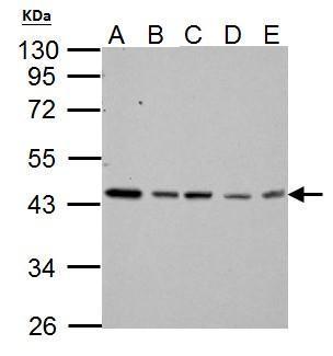
TARDBP antibody detects TARDBP protein by western blot analysis. A. 30 μg Neuro2A whole cell lysate/extract B. 30 μg GL261 whole cell lysate/extract C. 30 μg NIH-3T3 whole cell lysate/extract D. 30 μg BCL-1 whole cell lysate/extract E. 30 μg Raw264.7 whole cell lysate/extract 10% SDS-PAGE TARDBP antibody (GTX114210) dilution: 1:3000 The HRP-conjugated anti-rabbit IgG antibody (GTX213110-01) was used to detect the primary antibody. |
|
|
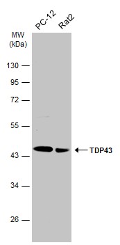
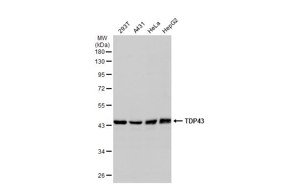
Various whole cell extracts (30 μg) were separated by 10% SDS-PAGE, and the membrane was blotted with TDP43 antibody (GTX114210) diluted at 1:2000. The HRP-conjugated anti-rabbit IgG antibody (GTX213110-01) was used to detect the primary antibody. |
|
|
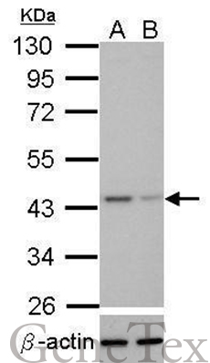
Western blot analysis of TDP-43 (GTX114210, upper panel) and beta-actin (GTX109639, lower panel)? Sample (30 μg of whole cell lysate) ? A: HeLa mock control? B: HeLa transfected shTDP-43 10% SDS PAGE? GTX114210 diluted at 1:1000. The HRP-conjugated anti-rabbit IgG antibody (GTX213110-01) was used to detect the primary antibody. |
|
|
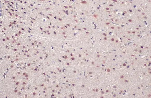
TDP43 antibody detects TDP43 protein at nucleus by immunohistochemical analysis.Sample: Paraffin-embedded mouse brain.TDP43 stained by TDP43 antibody (GTX114210) diluted at 1:500.Antigen Retrieval: Citrate buffer, pH 6.0, 15 min |
|
|
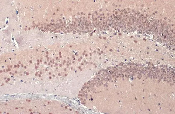
TDP43 antibody detects TDP43 protein at nucleus by immunohistochemical analysis.Sample: Paraffin-embedded rat brain.TDP43 stained by TDP43 antibody (GTX114210) diluted at 1:500.Antigen Retrieval: Citrate buffer, pH 6.0, 15 min |
|
|
Various whole cell extracts (30 μg) were separated by 10% SDS-PAGE, and the membrane was blotted with TDP43 antibody (GTX114210) diluted at 1:1000. The HRP-conjugated anti-rabbit IgG antibody (GTX213110-01) was used to detect the primary antibody. |
Produktgarantie und fachkundiger Support
