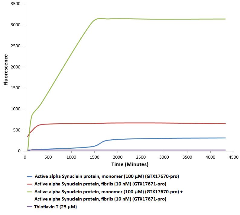
Endogenous alpha-synuclein phosphorylation. 100 µM alpha synuclein protein monomer (GTX17670-pro) seeded with 10 nM alpha synuclein protein PFF (GTX17671-pro) in 25 µM Thioflavin T (PBS pH 7.4, 100 µl reaction volume) generated an increased fluorescence
GTX17671-pro Image
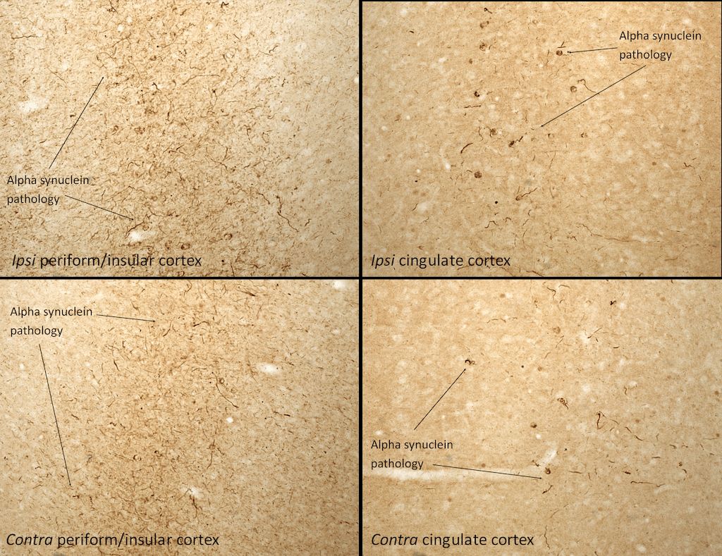
Immunohistochemistry analysis of rat brain injected with active mouse alpha synuclein PFFs (GTX17671-pro). Species: Female Sprague-Dawley Rat. Rat was injected with 2μL active mouse alpha synuclein PFFs (GTX17671-pro) in each of 2 injection sites: AP+1.6, ML+2.4, DV-4.2 from skull; and AP-1.4, ML+0.2, DV-2.8 from skull. 30 days post-injection. Fixation: Saline perfusion followed by 4% PFA fixation for 48 hrs. Secondary Antibody: Biotin-SP Donkey Anti-Rabbit IgG (H+L) at 1:500 for 2 hours in cold room with shaking. ABC signal amplification, DAB staining. Magnification: 20X. Alpha synuclein pathology is seen in the periform/insular cortex and the cingulate cortex on both the same (ipsi) and opposite (contra) sides as the injection sites.
Active alpha synuclein preformed fibrils (GTX17671-pro) seed the formation of new alpha synuclein fibrils from the pool of alpha synuclein monomers (GTX17670-pro). Thioflavin T is a fluorescent dye that binds to beta sheet-rich structures, such as those in alpha synuclein fibrils. Upon binding, the emission spectrum of the dye experiences a red-shift, and increased fluorescence intensity. Thioflavin T emission curves show increased fluorescence (correlated to alpha synuclein protein aggregation) over time when 10 nM of active alpha synuclein preformed fibrils (GTX17671-pro) is combined with 100 μM of alpha synuclein monomer (GTX17670-pro), as compared to active alpha synuclein preformed fibrils (GTX17671-pro) or alpha Synuclein monomer (GTX17670-pro) alone. Thioflavin T ex = 450 nm, em = 485 nm.
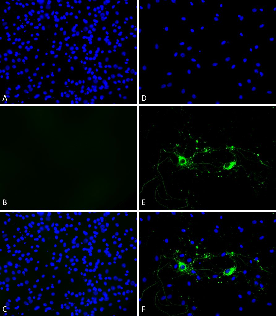
Primary rat hippocampal neurons (DIV16) show lewy body inclusion formation and loss of cells when treated with active mouse Alpha Synuclein Protein Preformed Fibrils (GTX17671-pro) at 4 μg/ml (D-F) on DVI2, but not when treated with a control (A-C). Tissue: Primary hippocampal neurons. Species: Sprague-Dawley rat. Fixation: 3% formaldehyde from PFA for 20 min. Blocker: 1:1 PBS:LiCOR Odyssey Block (LiCOR, 927-40010) and 30 mL/mL of 0.1% triton-X 100 for 30 min. Primary Antibody: Mouse anti-pSer129 Antibody (1:1000) and Rabbit anti-pSer129 (1:800) for 24 hours at 4ºC. Secondary Antibody: ATTO 546 Donkey Anti-Mouse (1:700) and ATTO 488 Donkey Anti-Rabbit (1:700) for 1 hour at RT (composite green). Counterstain: Hoechst (blue) nuclear stain at 1:3000 for 1 hour at RT. Localization: Lewy body incluscions. Magnification: 20x.
* Mehrwertsteuer und Versandkosten nicht enthalten. Irrtümer und Preisänderungen vorbehalten