
Collagen Hybridizing Peptide
Direct detection of unfolded collagen
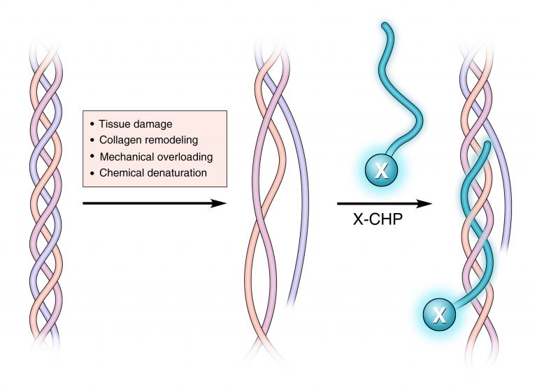
Collagen is the most abundant protein in the human body. It is a critical component of organs and tissues, providing the framework for cell attachment and growth. Collagen degradation regulates tissue remodeling, which occurs during development, homeostasis and repair, whereas excessive collagen degradation is observed in diseases such as cancer, inflammation, and fibrosis. All types of collagen share the triple helical protein structure, which is nearly exclusively found in collagen. After being cleaved by a collagenase, collagen molecule becomes thermally unstable at body temperature and the triple helix spontaneously denatures. Unfolding of the collagen triple helix also occurs during mechanical injuries of connective tissues and is an important damage mechanism.
.png) The founders of our partner 3Helix invented the Collagen Hybridizing Peptide (CHP), that can specifically bind to unfolded collagen molecules, by forming the triple helix with the denatured collagen chains, in a fashion analogous to a primer binding to a melted DNA strand during PCR. Conjugated with a fluorescent or biotin label, CHP is the first probe of its kind to enable direct detection of unfolded collagen molecules in virtually any tissue.
The founders of our partner 3Helix invented the Collagen Hybridizing Peptide (CHP), that can specifically bind to unfolded collagen molecules, by forming the triple helix with the denatured collagen chains, in a fashion analogous to a primer binding to a melted DNA strand during PCR. Conjugated with a fluorescent or biotin label, CHP is the first probe of its kind to enable direct detection of unfolded collagen molecules in virtually any tissue.

Applications of the Collagen Hybridizing Peptide (CHP)
Direct detection of collagen
CHP enables direct detection of unfolded collagen molecules in tissues, that have been subject to mechanical damage, or enzymatic remodeling (whether physiologically regulated, or disease associated).
Tissue decellularization
Collagen is one of the most widely used natural scaffold materials for regenerative medicine. The process of harvesting native extracellular matrices by removing cells from animal tissues (tissue decellularization) may alter the collagen structure and negatively affect the mechanical property and regenerative capacity of the ECM materials. CHP enables assessment of the structural integrity of collagen matrices from the molecular level, and facilitates optimization of the decellularization protocols.
Collagen identification
CHP can be used in several biochemical assays, such as in-gel Western blot, for identification and possibly quantification of collagen content in a biological sample.
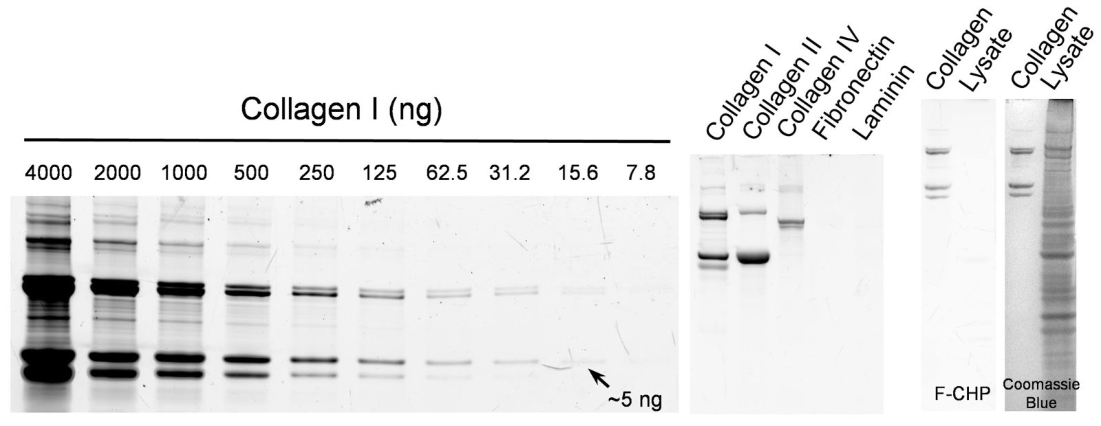
FIG: Fluorescent F-CHP reliably measures a dilution series of type I collagen directly in an SDS-PAGE gel, and detects a band containing as little as 5 ng of protein (left). CHP identifies multiple collagen types (middle) and has no affinity to non-collagenous molecules in the ECM (middle) and cell lysate (right) due to its neutral and hydrophilic nature.
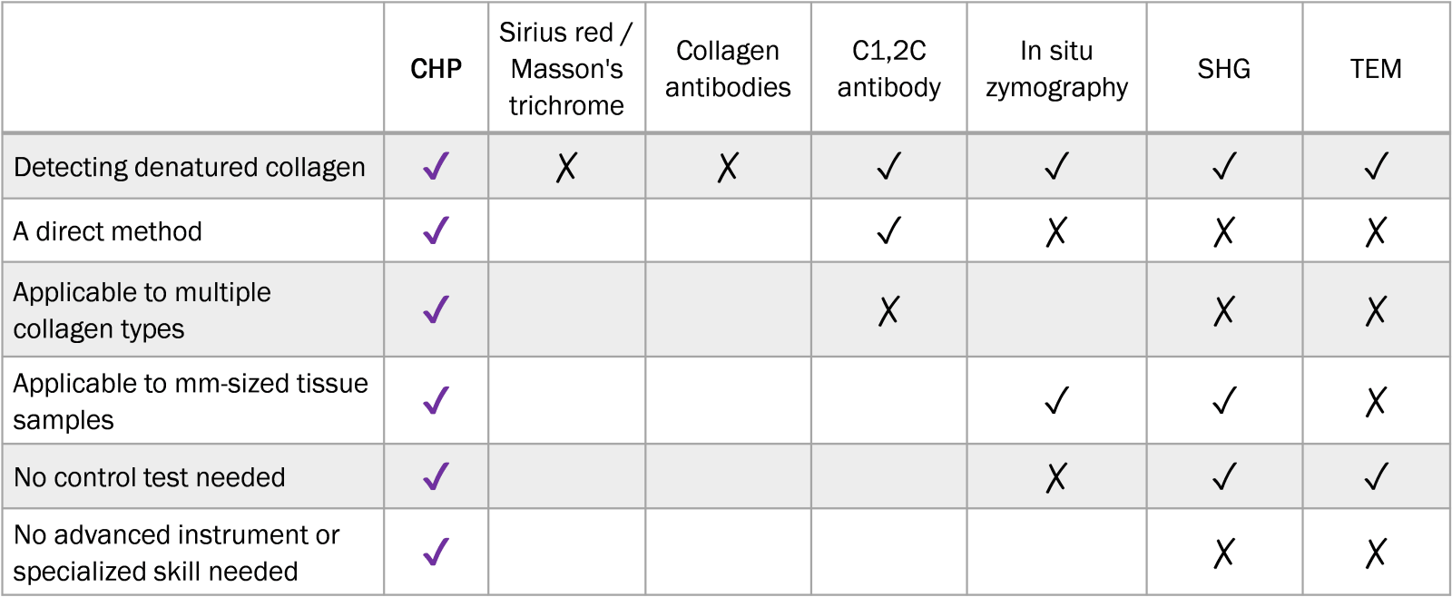
CHP versus conventional collagen characterization methods:
This might also interest you:
>> Webinar: Collagen Hybridizing Peptides
>> ELISA Kits for type I and type II collagen quantification
>> ScyTek Special Stains for Histology
09.02.2023

Labortops #4
Super Sale: up to 70% off labware

BIOZOL End-Of-Year P...
Up to 30 % discount on selected suppliers

iLite® Cell-Based So...
Reporter gene assays from Svar

Up to 50 % discount
Our current fall offers

Autumn conferences
Meet BIOZOL in Frankfurt, Bremen, and Düsseldorf