Recombinant Antibodies
Huabio Sample-Sized Rabbit Monoclonals
Recombinant antibodies have many advantages for the user, including unlimited availability of sequence-defined clones and cost-efficient production. Many methodological developments and findings in modern immunology would not have been possible without monoclonal antibodies from hybridomas.
Our partner Huabio offers a curated catalog of recombiant antibodies, created in-house with one mission: quality over quantity. Every product in Huabio's catalog is made and tested in-house, backed with the HUABIO guarantee.
Give it a try! Huabio is offering a selection of 20µL test size antibodies for you to determine which antibody is right for your experiment without a significant investment. So try out one (or five) Huabio antibodies today!
Highlighted Recombinant Rabbit Monoclonals:
Recombinant Active Caspase-3 Monoclonal Antibody (HBO-ET1602-47 / HBO-ET1602-47TR)
Huabio's peer-reviewed recombinant rabbit monoclonal primary has been validated for use with human and porcine samples in western blotting, immunohistochemistry, and immunocytochemistry.
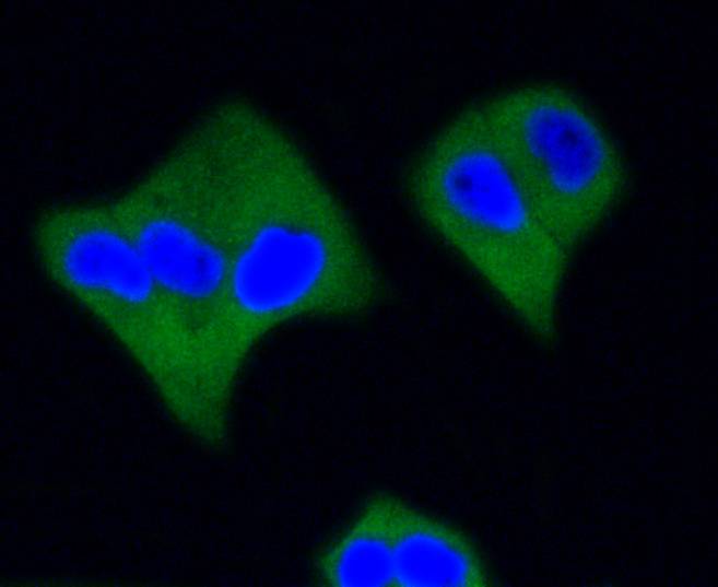 |
ICC staining of Active Caspase-3 in Hela cells (green). Formalin fixed cells were permeabilized with 0.1% Triton X-100 in TBS for 10 minutes at room temperature and blocked with 1% Blocker BSA for 15 minutes at room temperature. Cells were probed with the primary antibody (ET1602-47, 1/50) for 1 hour at room temperature, washed with PBS. Alexa Fluor®488 Goat anti-Rabbit IgG was used as the secondary antibody at 1/1,000 dilution. The nuclear counter stain is DAPI (blue). |
Recombinant PARP Monoclonal Antibody (HBO-ET1608-56 / HBO-ET1608-56TR)
This recombinant rabbit monoclonal primary antibody has been validated for use in western blotting, immunohistochemistry, immunocytochemsitry, and flow cytometry for human, mouse and rat samples.
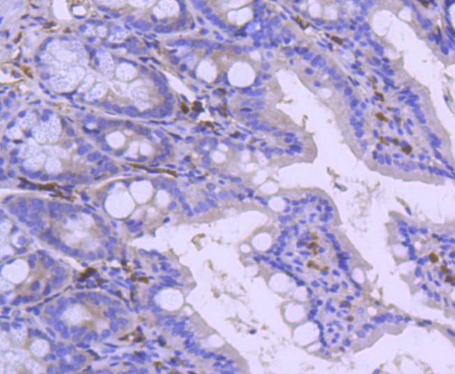 |
Immunohistochemical analysis of paraffin-embedded mouse colon tissue using anti-PARP antibody. The section was pre-treated using heat mediated antigen retrieval with Tris-EDTA buffer (pH 8.0-8.4) for 20 minutes.The tissues were blocked in 5% BSA for 30 minutes at room temperature, washed with ddH2O and PBS, and then probed with the primary antibody (ET1608-56, 1/50) for 30 minutes at room temperature. The detection was performed using an HRP conjugated compact polymer system. DAB was used as the chromogen. Tissues were counterstained with hematoxylin and mounted with DPX. |
Recombinant ATG5 Monoclonal Antibody (HBO-ET1611-38 / HBO-ET1611-38TR)
This peer-reviewed recombinant rabbit monoclonal is validated in human, mouse and rat samples for use in western blotting, immunohistochemistry, immunocytochemsitry, flow cytometry, and IP.
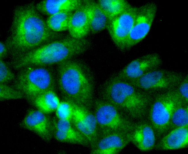 |
ICC staining of ATG5 in Hela cells (green). Formalin fixed cells were permeabilized with 0.1% Triton X-100 in TBS for 10 minutes at room temperature and blocked with 1% Blocker BSA for 15 minutes at room temperature. Cells were probed with the primary antibody (ET1611-38, 1/50) for 1 hour at room temperature, washed with PBS. Alexa Fluor®488 Goat anti-Rabbit IgG was used as the secondary antibody at 1/1,000 dilution. The nuclear counter stain is DAPI (blue) |
11.05.2021
Cell Manipulation
MDR Approval for Medical Devices
Labortops #2
Super Sale: up to 88% off labware
Obesity and Metaboli...
Research Reagents from Rockland, GeneTex and more

Spring Promo
Up to 30 % off Antibodies and ELISA kits

Protein Expression
MoBiTec Cloning and Expression Vectors

