
SOD1 antibody, Unconjugated, Rabbit, Polyclonal
Catalog Number:
GTX100554
- Images (9)
| Article Name: | SOD1 antibody, Unconjugated, Rabbit, Polyclonal |
| Biozol Catalog Number: | GTX100554 |
| Supplier Catalog Number: | GTX100554 |
| Alternative Catalog Number: | GTX100554-100,GTX100554-25 |
| Manufacturer: | GeneTex |
| Host: | Rabbit |
| Category: | Antikörper |
| Application: | ELISA, ICC, IHC-P, WB |
| Species Reactivity: | Aves, Human, Mouse, Rat, Reptile |
| Immunogen: | Recombinant protein encompassing a sequence within the center region of human SOD1. The exact sequence is proprietary. |
| Conjugation: | Unconjugated |
| Alternative Names: | superoxide dismutase 1 , ALS , ALS1 , HEL-S-44 , IPOA , SOD , hSod1 , homodimer |
| Application Notes: | WB: 1:500-1:10000. ICC/IF: 1:100-1:1000. IHC-P: 1:100-1:1000. ELISA: 1:1000-1:10000. *Optimal dilutions/concentrations should be determined by the researcher.Not tested in other applications. |
|
|
GTX100554 WB Image |
|
|
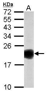
Sample (50 μg of whole cell lysate) A: mouse brain 15% SDS PAGE GTX100554 diluted at 1:1000 The HRP-conjugated anti-rabbit IgG antibody (GTX213110-01) was used to detect the primary antibody. |
|
|
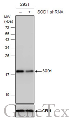
Non-transfected (–) and transfected (+) 293T whole cell extracts (30 μg) were separated by 15% SDS-PAGE, and the membrane was blotted with SOD1 antibody (GTX100554) diluted at 1:5000. The HRP-conjugated anti-rabbit IgG antibody (GTX213110-01) was used to detect the primary antibody. |
|
|
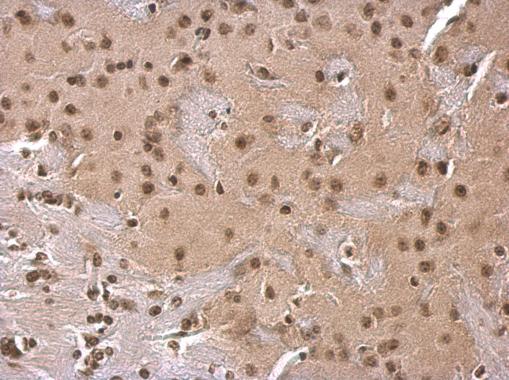
SOD1 antibody detects SOD1 protein at cytosol on mouse fore brain by immunohistochemical analysis. Sample: Paraffin-embedded mouse fore brain. SOD1 antibody (GTX100554) dilution: 1:500. Antigen Retrieval: Trilogy™ (EDTA based, pH 8.0) buffer, 15min |
|
|
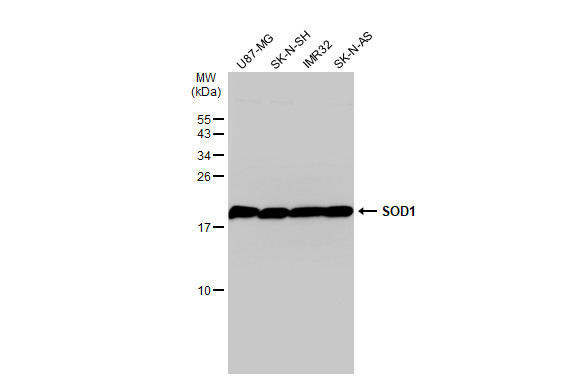
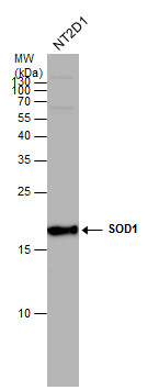
SOD1 antibody detects SOD1 protein by western blot analysis. Whole cell extracts (30 μg) was separated by 15% SDS-PAGE, and the membrane was blotted with SOD1 antibody (GTX100554) at a dilution of 1:1000. The HRP-conjugated anti-rabbit IgG antibody (GTX213110-01) was used to detect the primary antibody. |
|
|
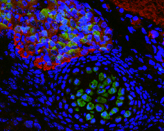
Immunofluorescence photomicrographs of paraffin-embedded mouse fetal brain. Green: SOD1 antibody (GTX100554) diluted at 1:200. The signal was developed using goat anti-rabbit IgG antibody (Dylight488) (GTX213110-04). Red: beta Tubulin 3/ TUJ1 antibody [GT11710] diluted at 1:100. The signal was developed using goat anti-mouse IgG antibody (Dylight594) (GTX213111-05). Blue: Nuclear staining with Hoechst 33342. Antigen Retrieval: Citrate buffer, pH 6.0, 15 min |
|
|
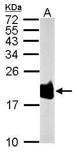
Sample (50 μg of whole cell lysate) A: Rat brain 15% SDS PAGE GTX100554 diluted at 1:1000 The HRP-conjugated anti-rabbit IgG antibody (GTX213110-01) was used to detect the primary antibody. |
|
|
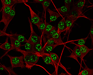
SOD1 antibody detects SOD1 protein at nucleus by immunofluorescent analysis. Sample: U-87 MG cells were fixed in 4% paraformaldehyde at RT for 15 min. Green: SOD1 protein stained by SOD1 antibody (GTX100554) diluted at 1:500. Red: beta Tubulin 3/ TUJ1 protein stained by beta Tubulin 3/ TUJ1 antibody (GTX631836) diluted at 1:200. |
|
|
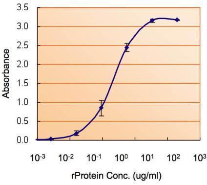
ELISA detection of SOD1 using GTX100554 for capture at a concentration of 5 μg/mL and GTX89049 for detection at a concentration of 1.5 μg/mL. |
Product Guarantee and Expert Support
