
Annexin A1 antibody, Unconjugated, Rabbit, Polyclonal
Catalog Number:
GTX101070
- Images (9)
| Article Name: | Annexin A1 antibody, Unconjugated, Rabbit, Polyclonal |
| Biozol Catalog Number: | GTX101070 |
| Supplier Catalog Number: | GTX101070 |
| Alternative Catalog Number: | GTX101070-100,GTX101070-25 |
| Manufacturer: | GeneTex |
| Host: | Rabbit |
| Category: | Antikörper |
| Application: | ICC, IHC-P, IP, WB |
| Species Reactivity: | Human, Mouse, Rat |
| Immunogen: | Recombinant protein encompassing a sequence within the center region of human Annexin A1. The exact sequence is proprietary. |
| Conjugation: | Unconjugated |
| Alternative Names: | annexin A1 , ANX1 , LPC1 |
| Application Notes: | WB: 1:500-1:3000. ICC/IF: 1:100-1:1000. IHC-P: 1:100-1:1000. IP: 1:100-1:500. *Optimal dilutions/concentrations should be determined by the researcher.Not tested in other applications. |
|
|
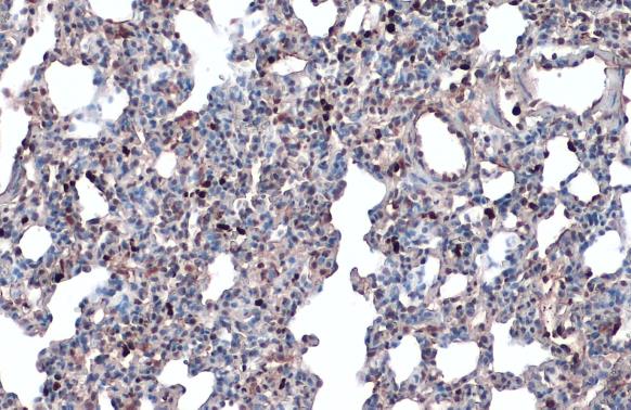
GTX101070 IHC-P Image |
|
|
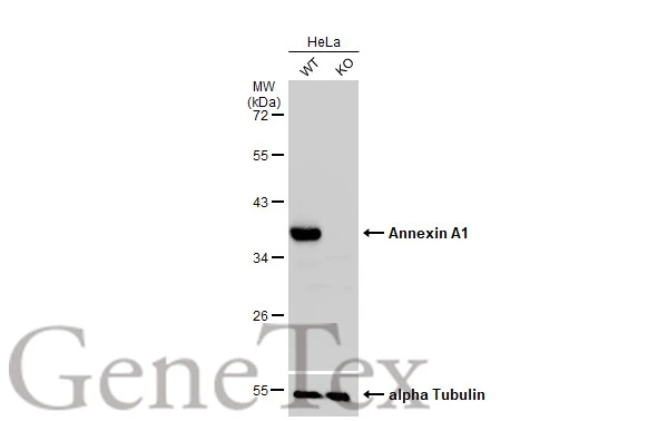
Wild-type (WT) and Annexin A1 knockout (KO) HeLa cell extracts (30 μg) were separated by 10% SDS-PAGE, and the membrane was blotted with Annexin A1 antibody (GTX101070) diluted at 1:1000. The HRP-conjugated anti-rabbit IgG antibody (GTX213110-01) was used to detect the primary antibody. |
|
|
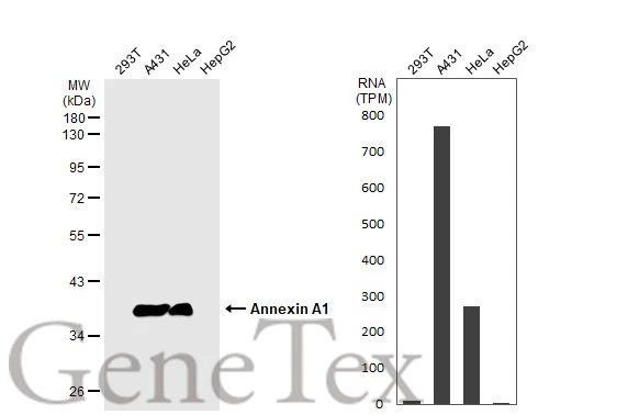
Various whole cell extracts (30 μg) were separated by 10% SDS-PAGE, and the membrane was blotted with Annexin A1 antibody (GTX101070) diluted at 1:10000. The HRP-conjugated anti-rabbit IgG antibody (GTX213110-01) was used to detect the primary antibody. Corresponding RNA expression data for the same cell lines are based on Human Protein Atlas program. |
|
|
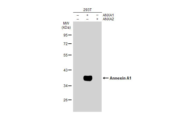
Non-transfected (–) and transfected (+) 293T whole cell extracts (30 μg) were separated by 10% SDS-PAGE, and the membrane was blotted with Annexin A1 antibody (GTX101070) diluted at 1:10000. The HRP-conjugated anti-rabbit IgG antibody (GTX213110-01) was used to detect the primary antibody. |
|
|
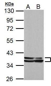
Sample (30 μg of whole cell lysate) A: NIH-3T3 B: JC 10% SDS PAGE GTX101070 diluted at 1:10000 The HRP-conjugated anti-rabbit IgG antibody (GTX213110-01) was used to detect the primary antibody. |
|
|
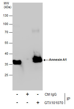
Immunoprecipitation of Annexin A1 protein from HeLa whole cell extracts using 5 μg of Annexin A1 antibody (GTX101070). Western blot analysis was performed using Annexin A1 antibody (GTX101070). EasyBlot anti-Rabbit IgG (GTX221666-01) was used as a secondary reagent. |
|
|
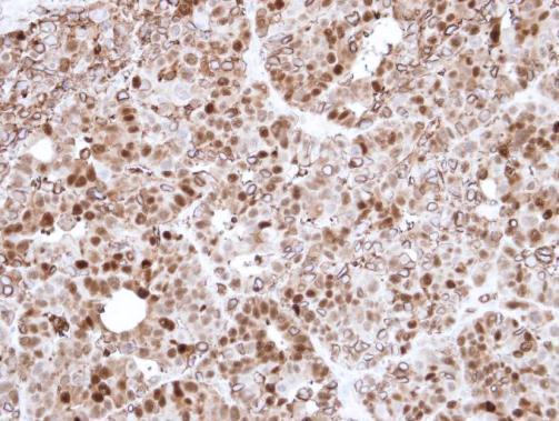
Immunohistochemical analysis of paraffin-embedded N87 Xenograft , using Annexin I(GTX101070) antibody at 1:500 dilution. Antigen Retrieval: Trilogy™ (EDTA based, pH 8.0) buffer, 15min |
|
|
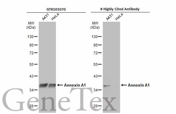
Various whole cell extracts (30 μg) were separated by 10% SDS-PAGE, and the membranes were blotted with Annexin A1 antibody (GTX101070) diluted at 1:5000 and competitor's antibody diluted at 1:5000. The HRP-conjugated anti-rabbit IgG antibody (GTX213110-01) was used to detect the primary antibody. *The competitor is not affiliated with GeneTex and does not endorse this product. |
|
|
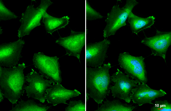
Annexin A1 antibody detects Annexin A1 protein by immunofluorescent analysis.Sample: HeLa cells were fixed in 4% paraformaldehyde at RT for 15 min.Green: Annexin A1 stained by Annexin A1 antibody (GTX101070) diluted at 1:500.Blue: Fluoroshield with DAPI (GTX30920).Scale bar= 10 μm. |
Product Guarantee and Expert Support
