
Calretinin antibody, Unconjugated, Rabbit, Polyclonal
Catalog Number:
GTX103261
- Images (9)
| Article Name: | Calretinin antibody, Unconjugated, Rabbit, Polyclonal |
| Biozol Catalog Number: | GTX103261 |
| Supplier Catalog Number: | GTX103261 |
| Alternative Catalog Number: | GTX103261-100,GTX103261-25 |
| Manufacturer: | GeneTex |
| Host: | Rabbit |
| Category: | Antikörper |
| Application: | ICC, IHC-Fr, IHC-P, WB |
| Species Reactivity: | Human, Mouse, Rat |
| Immunogen: | Recombinant protein encompassing a sequence within the center region of human Calretinin. The exact sequence is proprietary. |
| Conjugation: | Unconjugated |
| Alternative Names: | calbindin 2 , CAB29 , CAL2 , CR |
| Application Notes: | WB: 1:500-1:10000. ICC/IF: 1:100-1:1000. IHC-P: 1:100-1:1000. IHC-Fr: 1:100-1:1000. *Optimal dilutions/concentrations should be determined by the researcher.Not tested in other applications. |
|
|
GTX103261 WB Image |
|
|
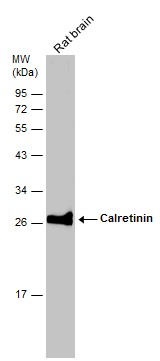
Rat tissue extract (50 μg) was separated by 12% SDS-PAGE, and the membrane was blotted with Calretinin antibody (GTX103261) diluted at 1:1000. The HRP-conjugated anti-rabbit IgG antibody (GTX213110-01) was used to detect the primary antibody. |
|
|
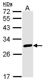
Sample (50 μg of whole cell lysate) A: mouse brain 12% SDS PAGE GTX103261 diluted at 1:1000 The HRP-conjugated anti-rabbit IgG antibody (GTX213110-01) was used to detect the primary antibody. |
|
|
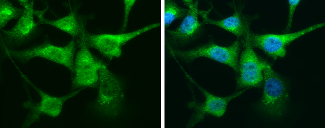
Calretinin antibody detects Calretinin protein at cytoplasm by immunofluorescent analysis. Sample: U87-MG cells were fixed in 4% paraformaldehyde at RT for 15 min. Green: Calretinin protein stained by Calretinin antibody (GTX103261) diluted at 1:200. Blue: Hoechst 33342 staining. |
|
|
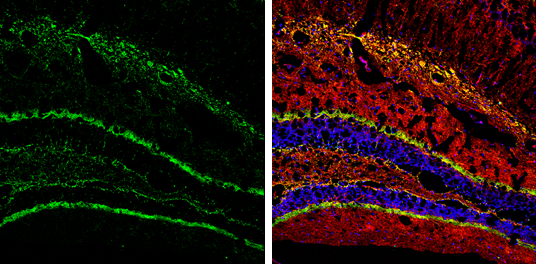
Calretinin antibody detects Calretinin protein by immunohistochemical analysis. Samples: Frozen Section adult mouse hippocampus. Green: Calretinin protein stained by Calretinin antibody (GTX103261) diluted at 1:250. Red: NF-H, stained by NF-H antibody [GT114] (GTX634289) diluted at 1:500. Blue: Fluoroshield with DAPI (GTX30920). |
|
|
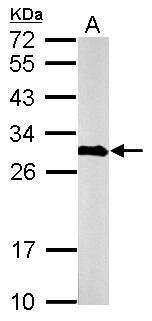
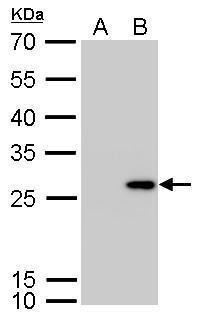
Calretinin antibody detects CALB2 protein by western blot analysis. A. 30 μg 293T whole cell lysate/extract B. 30 μg whole cell lysate/extract of human CALB2-transfected 293T cells 12% SDS-PAGE Calretinin antibody (GTX103261) dilution: 1:5000 The HRP-conjugated anti-rabbit IgG antibody (GTX213110-01) was used to detect the primary antibody. |
|
|
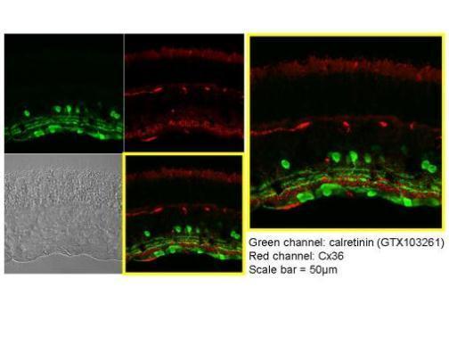
Immunohistochemical analysis of paraffin-embedded Mouse retina, using Calretinin(GTX103261) antibody at 1:250 dilution. Antigen Retrieval: Trilogy™ (EDTA based, pH 8.0) buffer, 15min |
|
|
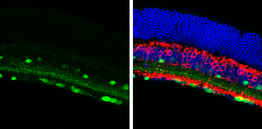
Calretinin antibody detects Calretinin protein in the amacrine cells by immunohistochemical analysis. Sample: Frozen sectioned adult mouse retina. Green: Calretinin protein stained by Calretinin antibody (GTX103261) diluted at 1:250. Red: Protein kinase C alpha staining. Blue: Fluoroshield with DAPI (GTX30920). |
|
|
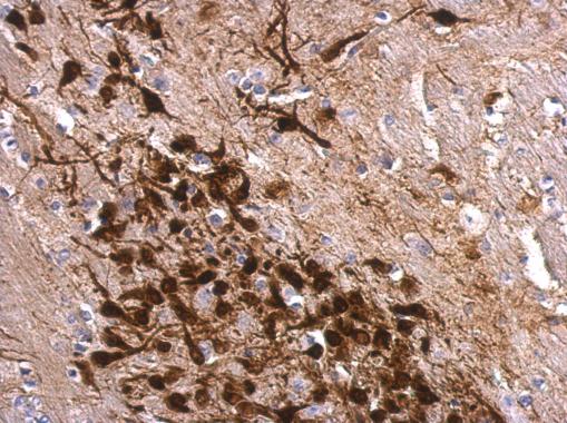
Calretinin antibody detects Calretinin protein at cytosol on mouse fore brain by immunohistochemical analysis. Sample: Paraffin-embedded mouse fore brain. Calretinin antibody (GTX103261) dilution: 1:500. Antigen Retrieval: Trilogy™ (EDTA based, pH 8.0) buffer, 15min |
Product Guarantee and Expert Support
