
SP1 antibody, Unconjugated, Rabbit, Polyclonal
Catalog Number:
GTX110593
- Images (9)
| Article Name: | SP1 antibody, Unconjugated, Rabbit, Polyclonal |
| Biozol Catalog Number: | GTX110593 |
| Supplier Catalog Number: | GTX110593 |
| Alternative Catalog Number: | GTX110593-100,GTX110593-25 |
| Manufacturer: | GeneTex |
| Host: | Rabbit |
| Category: | Antikörper |
| Application: | ChIP, ICC, IHC-P, IP, WB |
| Species Reactivity: | Human |
| Immunogen: | Recombinant protein encompassing a sequence within the center region of human SP1. The exact sequence is proprietary. |
| Conjugation: | Unconjugated |
| Alternative Names: | Sp1 transcription factor |
| Clonality: | Polyclonal |
| Concentration: | 0.22 mg/ml (Please refer to the vial label for the specific concentration.) |
| Molecular Weight: | 81 |
| Sensitivity: | We do not recommend use of this product for Mouse,Rat. |
| NCBI: | 6667 |
| UniProt: | P08047 |
| Buffer: | 1XPBS pH7, 1% BSA, 20% Glycerol, 0.025% ProClin 300. |
| Purity: | Purified by antigen-affinity chromatography. |
| Form: | Liquid |
| Application Notes: | WB: 1:500-1:3000. ICC/IF: 1:100-1:1000. IHC-P: 1:100-1:1000. IP: 1:100-1:500. *Optimal dilutions/concentrations should be determined by the researcher.Not tested in other applications. |
|
|
GTX110593 IHC-P Image |
|
|
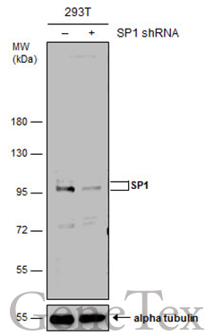
Non-transfected (–) and transfected (+) 293T whole cell extracts (50 μg) were separated by 7.5% SDS-PAGE, and the membrane was blotted with SP1 antibody (GTX110593) diluted at 1:10000. The HRP-conjugated anti-rabbit IgG antibody (GTX213110-01) was used to detect the primary antibody, and the signal was developed with Trident ECL plus-Enhanced. |
|
|
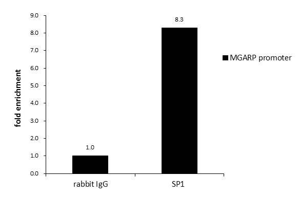
ChIP was performed with 293T chromatin extract and 5 μg of either normal rabbit IgG or anti-SP1 antibody. The precipitated DNA was detected by PCR with primer set targeting to MGARP promoter. |
|
|
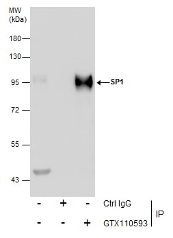
Immunoprecipitation of SP1 protein from THP-1 whole cell extracts using 5 μg of OCT1 antibody (GTX110593). Western blot analysis was performed using OCT1 antibody (GTX110593) diluted at 1:250. EasyBlot anti-Rabbit IgG (GTX221666-01) was used as a secondary reagent. |
|
|
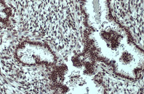
Immunohistochemical analysis of paraffin-embedded C2C12 xenograft, using SP1(GTX110593) antibody at 1:500 dilution. Antigen Retrieval: Trilogy™ (EDTA based, pH 8.0) buffer, 15min |
|
|
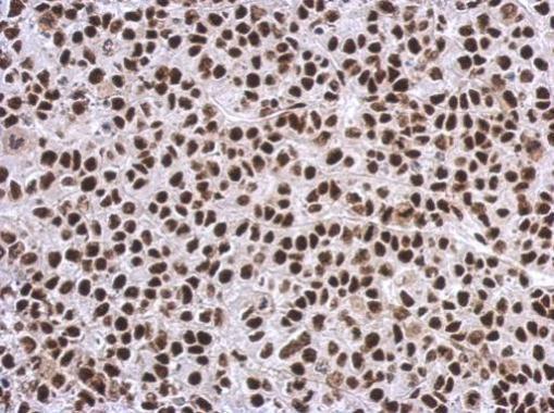
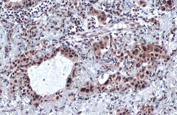
SP1 antibody detects SP1 protein at cytoplasm and nucleus by immunohistochemical analysis.Sample: Paraffin-embedded human lung cancer.SP1 stained by SP1 antibody (GTX110593) diluted at 1:2000.Antigen Retrieval: Citrate buffer, pH 6.0, 15 min |
|
|
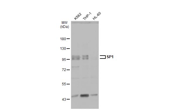
Various whole cell extracts (30 μg) were separated by 7.5% SDS-PAGE, and the membrane was blotted with SP1 antibody (GTX110593) diluted at 1:2000. The HRP-conjugated anti-rabbit IgG antibody (GTX213110-01) was used to detect the primary antibody. |
|
|
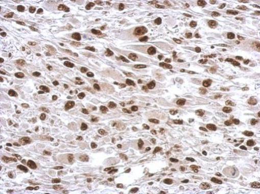
SP1 antibody detects SP1 protein at nucleus by immunohistochemical analysis.Sample: Paraffin-embedded human breast carcinoma.SP1 stained by SP1 antibody (GTX110593) diluted at 1:500.Antigen Retrieval: Citrate buffer, pH 6.0, 15 min |
|
|
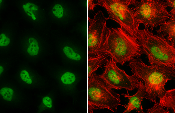
SP1 antibody detects SP1 protein at nucleus by immunofluorescent analysis.Sample: HeLa cells were fixed in 4% paraformaldehyde at RT for 15 min.Green: SP1 stained by SP1 antibody (GTX110593) diluted at 1:1000.Red: phalloidin, a cytoskeleton marker, diluted at 1:200. |
Product Guarantee and Expert Support
