
GSTP1 antibody, Unconjugated, Rabbit, Polyclonal
Catalog Number:
GTX112695
- Images (9)
| Article Name: | GSTP1 antibody, Unconjugated, Rabbit, Polyclonal |
| Biozol Catalog Number: | GTX112695 |
| Supplier Catalog Number: | GTX112695 |
| Alternative Catalog Number: | GTX112695-100,GTX112695-25 |
| Manufacturer: | GeneTex |
| Host: | Rabbit |
| Category: | Antikörper |
| Application: | ICC, IHC-Fr, IHC-P, WB |
| Species Reactivity: | Human, Mouse, Rat |
| Immunogen: | Recombinant protein encompassing a sequence within the center region of human GSTP1. The exact sequence is proprietary. |
| Conjugation: | Unconjugated |
| Alternative Names: | glutathione S-transferase pi 1 , DFN7 , FAEES3 , GST3 , GSTP , HEL-S-22 , PI |
| Application Notes: | WB: 1:500-1:10000. ICC/IF: 1:100-1:1000. IHC-P: 1:100-1:1000. *Optimal dilutions/concentrations should be determined by the researcher.Not tested in other applications. |
|
|
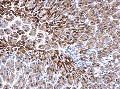
GTX112695 IHC-P Image |
|
|
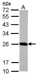
Sample (50 μg of whole cell lysate) A: mouse brain 12% SDS PAGE GTX112695 diluted at 1:1000 The HRP-conjugated anti-rabbit IgG antibody (GTX213110-01) was used to detect the primary antibody. |
|
|
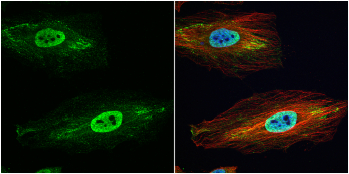
GSTP1 antibody detects GSTP1 protein at cytoplasm and nucleus by immunofluorescent analysis. Sample: HeLa cells were fixed in 4% paraformaldehyde at RT for 15 min. Green: GSTP1 protein stained by GSTP1 antibody (GTX112695) diluted at 1:500. Red: alpha Tubulin, a cytoskeleton marker, stained by alpha Tubulin antibody [GT114] (GTX628802) diluted at 1:1000. Blue: Hoechst 33342 staining. |
|
|
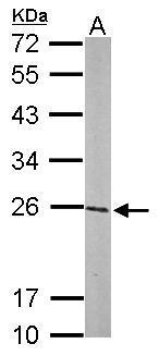
GSTP1 antibody detects GSTP1 protein by western blot analysis. A. 50 μg Rat brain lysate/extract 12% SDS-PAGE GSTP1 antibody (GTX112695) dilution: 1:1000 The HRP-conjugated anti-rabbit IgG antibody (GTX213110-01) was used to detect the primary antibody. |
|
|
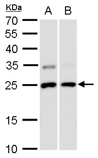
GSTP1 antibody detects GSTP1 protein by Western blot analysis. A. 30 μg A549 whole cell lysate/extract B. 30 μg HCT116 whole cell lysate/extract 12 % SDS-PAGE GSTP1 antibody (GTX112695) dilution: 1:5000 |
|
|
GSTP1 antibody detects GSTP1 protein at nucleus on rat fore brain by immunohistochemical analysis. Sample: Paraffin-embedded rat fore brain. GSTP1 antibody (GTX112695) dilution: 1:500. Antigen Retrieval: Trilogy™ (EDTA based, pH 8.0) buffer, 15min |
|
|
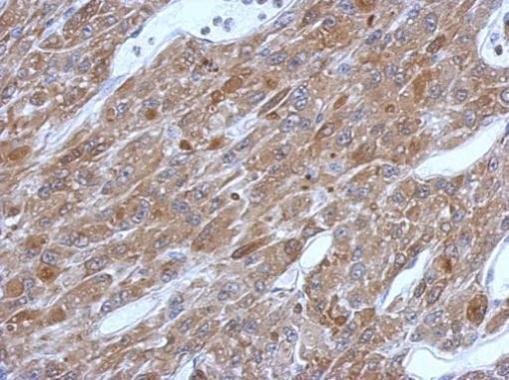
Immunohistochemical analysis of paraffin-embedded U87 xenograft, using GSTP1(GTX112695) antibody at 1:500 dilution. Antigen Retrieval: Trilogy™ (EDTA based, pH 8.0) buffer, 15min |
|
|
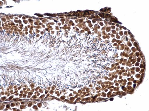
GSTP1 antibody detects GSTP1 protein at nucleus on mouse testis by immunohistochemical analysis. Sample: Paraffin-embedded mouse testis. GSTP1 antibody (GTX112695) dilution: 1:500. _x000D_ Antigen Retrieval: Trilogy™ (EDTA based, pH 8.0) buffer, 15min |
|
|
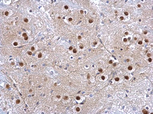
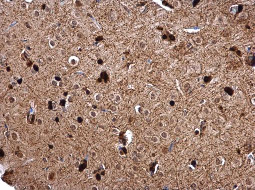
GSTP1 antibody detects GSTP1 protein at nucleus on mouse brain stem by immunohistochemical analysis. Sample: Paraffin-embedded mouse brain stem. GSTP1 antibody (GTX112695) dilution: 1:500. Antigen Retrieval: Trilogy™ (EDTA based, pH 8.0) buffer, 15min |
Product Guarantee and Expert Support
