
PLK1 antibody [13E8], IgG2b, Clone: [1.30E+09], Unconjugated, Mouse, Monoclonal
Catalog Number:
GTX15779
- Images (9)
| Article Name: | PLK1 antibody [13E8], IgG2b, Clone: [1.30E+09], Unconjugated, Mouse, Monoclonal |
| Biozol Catalog Number: | GTX15779 |
| Supplier Catalog Number: | GTX15779 |
| Alternative Catalog Number: | GTX15779-100 |
| Manufacturer: | GeneTex |
| Host: | Mouse |
| Category: | Antikörper |
| Application: | ELISA, ICC, IHC, IHC-P, IP, WB |
| Species Reactivity: | Human, Mouse, Rat |
| Immunogen: | Recombinant protein containing residues 300-400 of human PLK1. |
| Conjugation: | Unconjugated |
| Alternative Names: | polo like kinase 1 , PLK , STPK13 |
| The Ser/Thr protein kinase encoded by this gene belongs to the CDC5/Polo subfamily. It is highly expressed during mitosis and elevated levels are found in many different types of cancer. Depletion of this protein in cancer cells dramatically inhibited ce |
| Clonality: | Monoclonal |
| Concentration: | 1 mg/ml (Please refer to the vial label for the specific concentration.) |
| Clone Designation: | [1.30E+09] |
| Molecular Weight: | 68 |
| Isotype: | IgG2b |
| NCBI: | 5347 |
| UniProt: | P53350 |
| Buffer: | PBS, 1mg/ml BSA, 0.05% Sodium azide. |
| Source: | Human |
| Purity: | Protein G purified |
| Form: | Liquid |
| Application Notes: | WB: 1:1,000. ICC/IF: 3 µg/ml. IHC-P: 1:20. *Optimal dilutions/concentrations should be determined by the researcher.Not tested in other applications. |
|
|
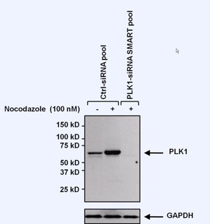
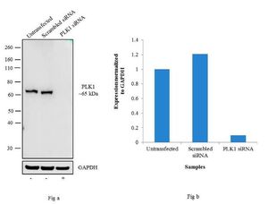
WB analysis of untreated or Nocodazole (100nM, 48 hours) treated U2OS lysate from non-targeting control or PLK1 siRNA transfected U2OS cells using GTX15779 PLK1 antibody [13E8]. Dilution : 1:1000 Loading : 25 μg |
|
|
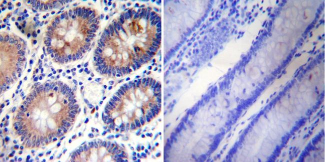
IHC-P analysis of human colon tissue using GTX15779 PLK1 antibody [13E8]. Left : Primary antibody Right : Negative control without primary antibody Antigen retrieval : heat induced antigen retrieval was performed using 10mM sodium citrate (pH6.0) buffer, microwaved for 8-15 minutes Dilution : 1:200 |
|
|
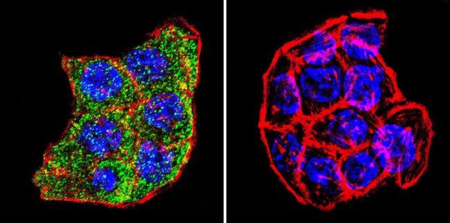
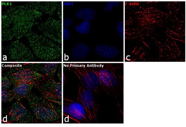
ICC/IF analysis of U251 cells using GTX15779 PLK1 antibody [13E8]. Cells were probed without (right) or with(left) an antibody. Green : Primary antibody Blue : Nuclei Red : Actin Fixation : formaldehyde Dilution : 1:20 overnight at 4ºC |
|
|
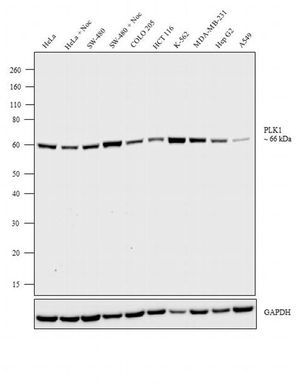
GTX15779 WB Image |
|
|
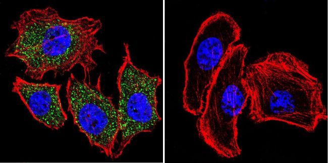
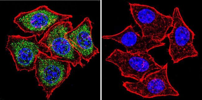
ICC/IF analysis of HeLa cells using GTX15779 PLK1 antibody [13E8]. Cells were probed without (right) or with(left) an antibody. Green : Primary antibody Blue : Nuclei Red : Actin Fixation : formaldehyde Dilution : 1:20 overnight at 4ºC |
|
|
WB analysis of PLK1 knockdown HeLa cells (lane 3), scrambled siRNA transfected cells (lane 2) and untransfected cells (lane 1) using GTX15779 PLK1 antibody [13E8]. Dilution : 1 μg/ml |
|
|
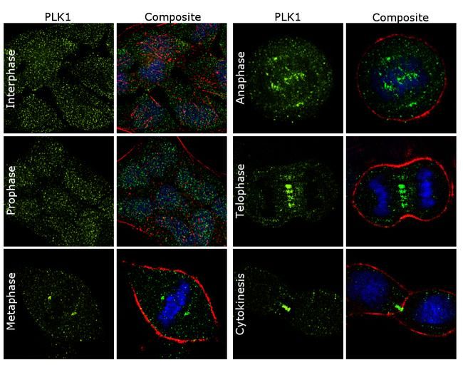
ICC/IF analysis of HeLa cells using GTX15779 PLK1 antibody [13E8]. Progression of cells through subsequent phases of the cell cycle results in differential localization of PLK1 in spindle poles, kinetochores and cleavage furrow. Green : Primary antibody Blue : Nuclei Red : Actin Fixation : 4% paraformaldehyde Permeabilization : 0.1% Triton X-100 for 10 minute Dilution : 3 μg/ml in 0.1% BSA and incubated for overnight at 4ºC |
|
|
ICC/IF analysis of WiDr cells using GTX15779 PLK1 antibody [13E8]. Cells were probed without (right) or with(left) an antibody. Green : Primary antibody Blue : Nuclei Red : Actin Fixation : formaldehyde Dilution : 1:20 overnight at 4ºC |
|
|
ICC/IF analysis of HeLa cells using GTX15779 PLK1 antibody [13E8]. Fixation : 4% paraformaldehyde Permeabilization : 0.1% Triton X-100 for 10 minute Dilution : 3 μg/ml in 0.1% BSA and incubated for overnight at 4ºC |
Product Guarantee and Expert Support
