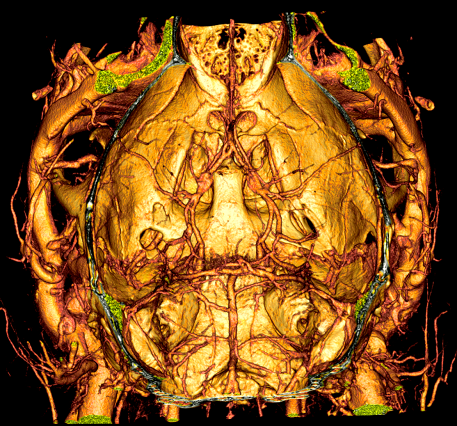
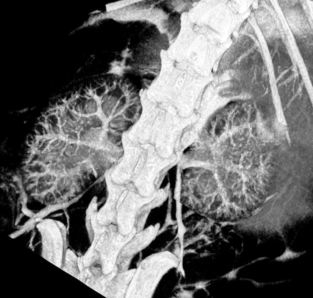
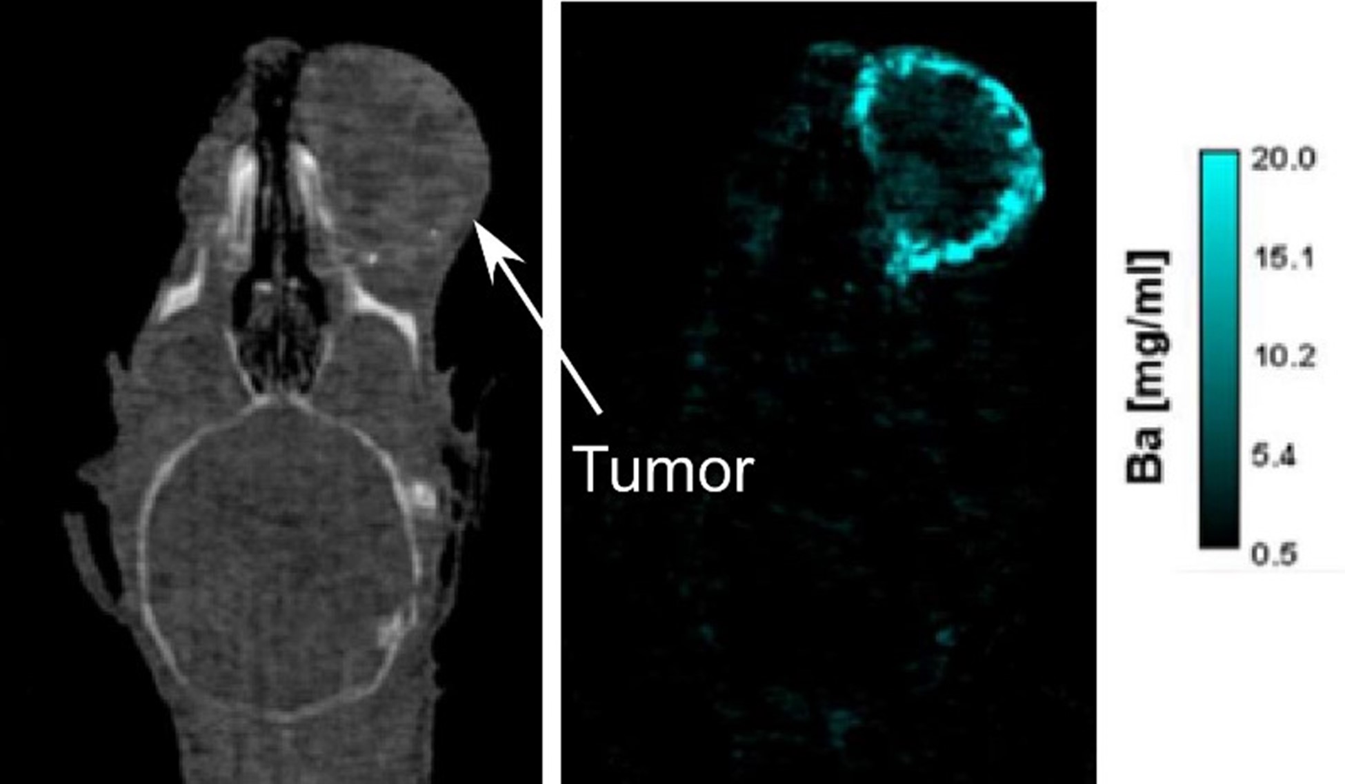
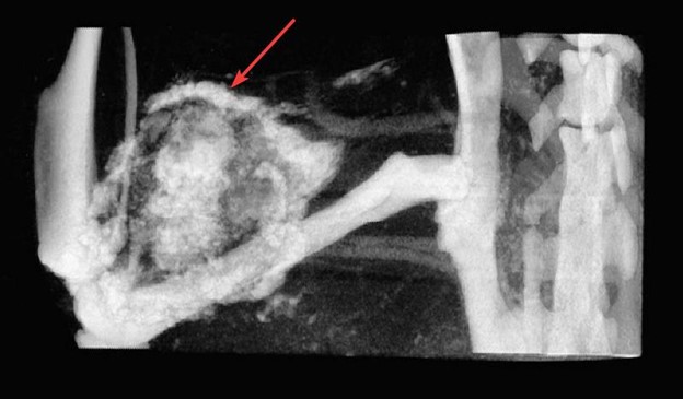
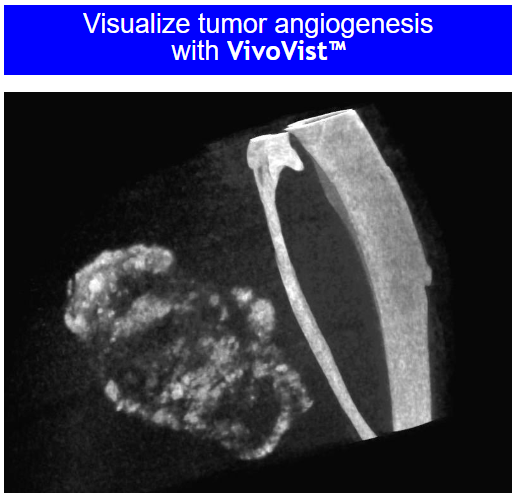
VivoVist™ is a revolutionary nanotechnology super contrast agent of alkaline earth metal nanoparticles. VivoVist™ enables greatly enhanced X-ray imaging of blood vessels, tumors, and other tissues and organs. It is particularly useful for in vivo live animal microCT imaging, for studies of tumors, stroke, atherosclerosis and other vascular conditions, organ function, and other biological structural and functional analyses.
Features and Advantages
- VivoVist™ provides approximately 3-4 times higher contrast than competing commercial microCT contrast agents. An Initial blood concentration of 50 mg/mL (5%), or 1 g/kg body weight (one-third of one vial for a 25 g mouse) gives greater than 3500 HU (Hounsfeld Units) contrast.
- Long blood half-life of 14 hrs. VivoVist™ stays in the circulatory system longer than competing products. This enables extended imaging times and data collection, and allows longer diffusion into tumors and other features of interest, increasing their contrast.
- Lowest price - makes imaging rats and larger animals affordable. VivoVist™ is more concentrated that competing products, meaning that fewer units are required to image larger animals. Less than two vials will provide good contrast in a 250 g rat.
- Low toxicity (4 g/kg is well-tolerated). VivoVist™ can be administered in higher doses than competing products, to produce super-high contrst in fine structures or to accelerate loading of tumors or other features of interest.
- Low osmolality, even at high concentrations. This means that injecting VivoVist™ will not greatly change the levels of electrolytes in the circulatory system. Impacts to metabolism, signaling and other processes that depend on maintaining a steady ionic strength are minimized.
- Low viscosity: easy to inject into small mouse tail vein blood vessels (typical injection volume 0.25 mL). VivoVist™ is more easily injected, causes less injection site trauma to the animal, and may be administered more quickly and in higher concentration that more viscous contrast agents such as iodine-based reagents.
- Can be imaged using MicroCT, clinical CT, planar X-ray, or mammography units. VivoVist™ is adaptable for any commonly used computed tomography system, giving users the flexibility to image in any desired instrument configuration.
- Enhances radiotherapy X-ray dose to tumors and other targets. Because it absorbs X-rays so strongly, VivoVist™ increases the local X-ray dose. If it is concentrated in tumors or other therapeutic targets, it can provide a method for enhancing radiotherapy and increasing the efficacy of cancer therapy.
|


